The history of X-rays dates back to the late 19th century. Here is a brief overview of the key milestones:
Discovery of X-rays: In 1895, Wilhelm Conrad Roentgen, a German physicist, accidentally discovered X-rays while experimenting with cathode rays. He noticed that a fluorescent screen in his lab started to glow even when it was not in direct contact with the cathode rays. Roentgen named these mysterious rays “X-rays” due to their unknown nature.
Early experiments: Roentgen conducted further experiments to study the properties of X-rays. He found that they could pass through various substances, including human tissue, but were absorbed by denser materials like bones and metal. This property made X-rays useful for medical imaging.
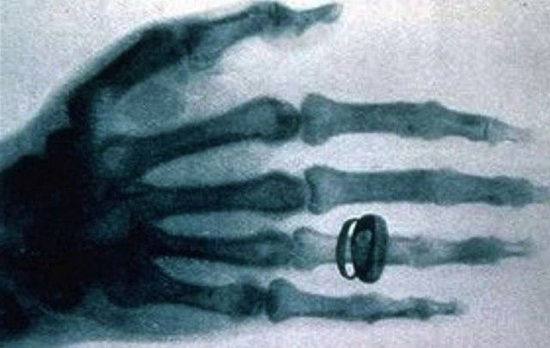
Medical applications: X-rays quickly found applications in the medical field. In 1896, Roentgen produced the first X-ray image of his wife’s hand, revealing the bones and a ring on her finger. This breakthrough led to the use of X-rays for diagnosing fractures, tumors, and other internal conditions.
Advancements in technology: Over the years, advancements in X-ray technology improved the quality and safety of imaging. In the early 20th century, X-ray machines became more portable and easier to use. Techniques like fluoroscopy, which allowed real-time X-ray imaging, were developed.
World War I and medical advancements: During World War I, X-rays played a crucial role in diagnosing and treating injuries. Portable X-ray units were used on the battlefield to locate bullets and fractures. This experience led to further advancements in X-ray technology and its applications.
Development of X-ray machines: In the 20th century, X-ray machines became more sophisticated. X-ray tubes were developed to generate higher-energy X-rays, enabling better penetration and imaging of dense tissues. Film-based X-ray imaging was the standard until the digital revolution in the late 20th century.
cigarette


